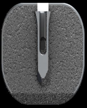
Overhead view of prodisc C, showing keel's tapered shape which facilitates implantation and fixation.
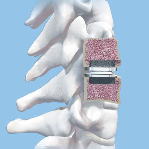
Illustration showing lateral view illustrating bone cuts to provide proper device fixation for prodisc C.
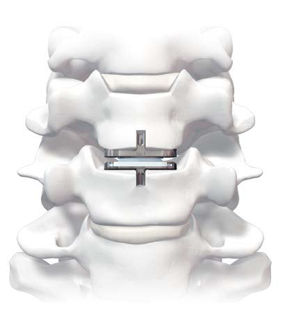
Illustration showing prodisc C anterior view illustrating keel fixation within the vertebral body.
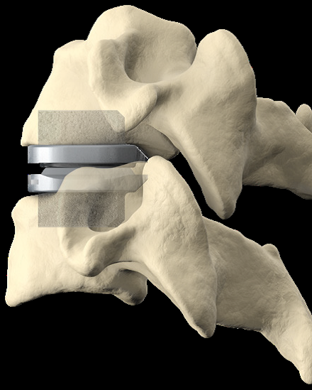
Illustration showing prodisc C from a lateral viewpoint within a bone model.
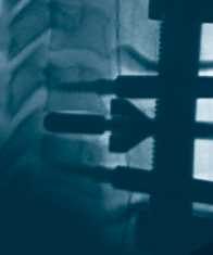
X-Ray showing lateral view with trial in place during a prodisc C implantation.
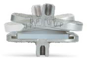
Image showing prodisc C as it goes from lateral flexion to lateral extension.
Available Search Tags
Watch this short primer on Centinel Spine and its unique and extraordinary place as a catalyst of change in the spine industry—with pioneering technologies and a clinical history that have led to successes ranging from PGA champions to a growing list of surgeon-patients.
 SEE MORE VIDEOS